|
The optical microscopes are
limited in their resolution R because it depends of the
wavelength according to the following relation:

with :
n : index of the medium
U: angle of the electron beam (half great point angle)
0,61: coefficient related to the diffraction of Fraunhofer.
 : is the wavelength of
the radiation
: is the wavelength of
the radiation
 for a radiation
of particle of mass m and speed v. for a radiation
of particle of mass m and speed v.
h : Planck's constant
An electron beam has a wavelength much lower than that of a beam
of light from where a resolution much smaller.
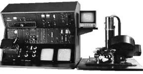
 The electronic transmission
miscroscope Siemens in the Museum
The electronic transmission
miscroscope Siemens in the Museum
Moreover, if the electron speed
is raised, the wavelength decreases. One obtains practical resolutions
provided in the table below.
|
(photon)
= 400 à 700 nm |
(electron)
= 0,001 nm |
|
R (photons) = 500 nm |
R (electron) = 0,2 nm |
|
1 nm =  m m
We can note that the value
of the resolution practises electron microscope is not that awaited
theoretically because the aperture of the electron beam cannot
exceed 1° contrary to the beam of light whose angle u
can reach 65°.
We can have a gain of resolution with the electronic microscopes
of 1000 X instead of 100 000 X in theory.
Essential qualities of an
electron microscope:
- to have a monocinetic beam
i.e. which one must be able to regulate the accelerating tension
with a 1 V precision.
- good lenses.
- for the electronic microscopes with transmission, a good factor
of penetration of the radiation which must be without damage
for the object observed.
|
The
transmission electron microscopy and the scanning electron microscopy |
|
The transmission electron microscopy principle is closed to the optical
microscope. The electronic beam goes throught the sample and
loses in the passing a certain number of electrons. The first
images were obtained for the first time by Ernst Ruska in 1931.
Principle :
 The
electron emission is produced by heating of a tungsten filament
or a crystal of lanthanum hexaborure. The
electron emission is produced by heating of a tungsten filament
or a crystal of lanthanum hexaborure.
A high vacuum is carried out
in the tube of the microscope.
The accelerating tension is
about 200 kV for the cheapest apparatuses and 1000 kV for most
expensive (2 $ per volt) !
The magnetic lenses made up
of a coil and an iron core, focus the electron beam. The variation
of the focal distance makes it possible to vary the enlargement
(up to 1 000 000 X) and the focus . The visual observations are
always relayed by a photograph catch. It is enough to make rock
the screen so that the photographic plates are impressed. The
focus does not need to be changed bus being given the low value
of U, the depth of field is very high. Practically, it
is necessary to use objects small thickness (0,5 nm) in order
to be as much as possible transparent to the electrons.
Moreover, the biological samples
must be dehydrated if not water present in these samples would
vaporize immediately being given the very low pressure reigning
in the emptied tube of its air.
Analyse d'une image de microscope
électronique à transmission

These photography shows an
enlarged yeast cell
20 000 X which divides by budding.
We observe a cut of the core,
while in the cytoplasm are present secretary organoids, which
make it possible the cell to secrete proteins in the external
medium.
Image given by Mrs. Morin-Ganet Doctor of biology which worked
on the yeast Saccharomyces cerevisiae in electron microscopy.
 The
scanning
electron microscope carried
out by Manfred von Ardenne in 1939 is an apparatus which balie
the sample of an electron beam. These electrons strike the sample
which emits in its turn of the secondary electrons of which the
number depends on nature on studied surface. The
scanning
electron microscope carried
out by Manfred von Ardenne in 1939 is an apparatus which balie
the sample of an electron beam. These electrons strike the sample
which emits in its turn of the secondary electrons of which the
number depends on nature on studied surface.
These are the electrons which are collected and detected. In
the case of the apparatus opposite, the enlargement can vary
from 10 X with 50 000 X and its resolution can reach 7 nm.
Scanning electron microscope
ETEC Autoscan (© 1995, ARS)
|
Optical
microscope |
Electron
microscope |
- light beam
- optical lens
- resolution : 500 nm
|
- electron beam
- electromagnetic lens
- resolution : 0,2 nm
|
|
1 nm = m
>> Site
map << |


